

Description
We provide first class ophthalmological services with the highest standars and latest technology, based in Goiania, the capital of the state of Goiás.<br><br>Our services include:<br> Adjustment of Contact Lenses: To have security in the use of contact lenses, it is essential that is well adapted, with curvature, diameter, grade and material suitable for each case.<br><br> Analyzer nerve fibers: The analyzer of nerve fibers is revolutionizing the propaedeutic of patients or suspected patients with glaucoma, as it quantifies, document and compares with confocal laser precision, the minimum changes in nerve fibers affected by glaucoma. Examination currently needed for patients or suspected patients with glaucoma.<br><br> Fluorescein angiography and digital: We were the first to carry out the examination of fluorescent angiography digital in Brazil. This is a technical investigation that is to be injecting a contrast (fluorescein sodium) into a vein of previous arm or the hand to see and photograph its way in vessels of the eye, as well as level of the retina and choroid. The images are analyzed by medical ophthalmologist with the aid of a computer. It is a great aid in the diagnosis of diseases reticoroideas (vascular, inflammatory and degenerative).<br><br> Biometrics: Biometrics uses A-mode ultrasound scan to get the measure of axial length of the eyeball and calculate the degree of artificial crystalline lens (intraocular lens) to be implanted during cataract surgery. It also serves for the diagnosis and monitoring of diseases that increase the size of the eyeball, such as congenital glaucoma. <br><br> Perimetry computed: The Computer Perimetry assesses the defects of the visual field and scotomas caused by certain diseases. indications are: Glaucoma (early diagnosis and monitor the evolution of the disease), Endocrinopathy, Maculopathy, and Neuropathies.<br><br> Keratometry computed: Keratometry Computer is an examination conducted by computer and measured the curvature of the corneal surface. This assessment is essential for adaptation of contact lenses and lens calculation of the artificial (intraocular lens) to be implanted during cataract surgery.<br><br> Study of stereoscopic optical disk: The study of stereoscopic Optical Disk is a means of permanent and detailed documentation of the optic disc, through photography, which are analyzed: ring neuro-retinal, C / D, depth of the excavation, displacement of retinal vessels, among other features. This is accomplished with the aid of IMAGENET.<br><br> OCT - optical coherence tomography: Optical coherence tomography (OCT) is a new test capable of making cuts of Sectional biological tissues. Its principle is very much that of B mode ultrasound, but instead of using sound waves makes use of light to obtain the images, which greatly increases its spatial resolution.<br><br> Paquimetria ultrasound: The Paquimetria Ultrasonic examination is one that accurately measured the thickness of the cornea.<br><br> Refraction computed: The refraction is a computerized test that checks the necessity or otherwise of the use of corrective lenses to improve vision. Where is achieved by a computer, independently of patient information. <br><br> Retinography color: The retinography Color is an examination in which photographed the bottom of the eye for documentation. <br><br> Tonometry: Tonometry is the measurement of intraocular pressure. Among the existing methods, the most reliable is the applanation tonometry, which is the prototype of tonômetro Goldmann. This type of tonometers measures the force needed to smooth over a standard area of corneal surface. <br><br> A and B ultrasound scan: There are two modes of Ultrasonography: (1) tThe so-Scan, is a dimensional representation of echoes, which provide characteristics of structures found; and (2) Mode B-Scan, is a two-dimensional representation of echoes, which provide topographical and anatomical characteristics of the region examined. <br><br> Videoangiography with digital indocyanine green: The Digital videoangiography with indocyanine green revolutionized the diagnosis of various diseases of fund-eye. So there are limits to this technique. Bleeding, pigments and fluids serosanguinolentos can block the view of fluorescein. It was there that appeared to videoagiografia digital computed, which is a technique of examination which consists of injecting a contrast (indocyanine green) into a vein of the forearm or hand to see and photograph its way to the bottom of the eye even if there are changes that could prevent the his view. It is necessary for carrying out this test a computerized system for digital images (IMAGEnet 640 or 1024) to produce images with high resolution.
- eye
More about
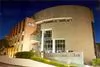
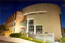
Centro Brasileiro De Cirurgia De Olhos
Not informed
Not informed
Not informed
Year
Established
Business type
Keywords
- eye surgery
Contact and location
-
 Marcos ********
-
+55 62********
-
Goiânia / GO | Brazil
